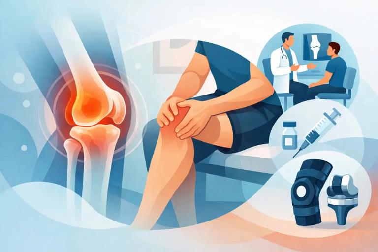

When shoulder pain stops you reaching overhead, or a swollen knee is affecting every step, waiting weeks without a clear answer can be frustrating. Understanding how point of care ultrasound works can help explain why it is such a useful part of a rapid-access musculoskeletal assessment. It allows a clinician to assess many soft-tissue structures in real time, discuss what they can see and, where appropriate, plan treatment within the same appointment.
Point-of-care ultrasound is performed by the clinician looking after you, rather than being arranged as a separate imaging appointment. For many tendon, joint and soft-tissue concerns, it can provide useful information immediately alongside your symptoms, examination findings and medical history.
How point of care ultrasound works
Ultrasound uses high-frequency sound waves to create images of structures beneath the skin. A small handheld device called a probe, or transducer, sends these sound waves into the body. Different tissues reflect the waves back at different speeds and strengths. The ultrasound machine converts those returning echoes into moving images on a screen.
Gel is placed on the skin before scanning. This removes the tiny pockets of air that would otherwise sit between the probe and skin and interfere with the sound waves. The gel can feel cool, but the scan itself does not use radiation and is not painful.
Unlike an X-ray, which is particularly useful for looking at bone, ultrasound is often well suited to superficial soft tissues. This includes tendons, ligaments, muscles, bursae, fluid around joints, cysts and some nerve-related problems. It can also be used to assess the surface of a joint and identify inflammation or changes in nearby soft tissues.
The images are live rather than static. Your clinician may ask you to move your shoulder, bend your knee, rotate your wrist or point your foot while scanning. This dynamic assessment can be especially valuable when symptoms occur only during movement. For example, a tendon may appear relatively normal when the arm is at rest but demonstrate irritation, thickening or altered movement in the position that reproduces pain.
What the clinician is looking for
The purpose of a scan is not simply to find an abnormality. Many people, particularly as they get older or remain active in sport, have changes on imaging that may not be the cause of their symptoms. Our clinicians can interpret the ultrasound images but more importantly match these to your clinical symptoms to make sure any changes are relevant and likely to be causative of your symptoms.
Depending on the body area and concern, ultrasound may identify tendon thickening or tearing, inflammation around a tendon or nerve, bursitis, joint effusion, synovitis, muscle injury, ganglion cysts, plantar fascia changes or features of osteoarthritis close to the joint surface. Colour Doppler may also be used. This displays blood flow and can help identify increased vascular activity associated with inflammation.
The unaffected side is sometimes scanned for comparison. That can be helpful where an image finding is subtle, or where normal anatomy varies from person to person.
Why real-time scanning can improve your appointment
In a conventional pathway, a patient may first see one clinician, wait for an imaging referral, attend a scan elsewhere and then wait again to discuss the result. Point-of-care ultrasound can reduce this fragmentation for suitable musculoskeletal problems. The person assessing your symptoms is also able to scan the painful area, test movement and explain the findings in context.
This does not mean every cause of pain can be diagnosed with ultrasound alone. It means the information is available at the point when clinical decisions are being made. That may give greater confidence to begin a targeted rehabilitation plan, arrange further imaging, refer for another opinion or consider an image-guided procedure.
For patients, the practical benefit is often clarity. Instead of leaving an appointment uncertain about what happens next, you can have a clearer explanation of the likely diagnosis, the structures involved and the most appropriate non-surgical options.
At Clinic 360, point-of-care ultrasound forms part of a specialist-led assessment for appropriate presentations. The scan is interpreted alongside your clinical examination rather than treated as an isolated test result. This approach is particularly useful for people seeking prompt answers for shoulder, elbow, hand, wrist, hip, knee, ankle and foot symptoms.
What happens during a musculoskeletal ultrasound scan?
A point-of-care scan is usually straightforward. You will be positioned so that the relevant area is easy to examine, which may mean sitting or lying down. Clothing may need to be moved away from the area, but you should only need to expose the body part being assessed.
Your clinician applies gel and moves the probe across the skin in several directions. They may apply light pressure or ask you to move through a particular range. If an area is tender, let them know. While some pressure may be necessary to obtain a clear image, the examination should be adapted where possible to keep you comfortable.
A focused scan can often be completed within the consultation, although the time required depends on the problem and body area. The findings should be explained clearly, including what the scan can show, what remains uncertain and whether another test would add useful information. A documented report after our appointment will always be provided.
When ultrasound-guided treatment may be considered
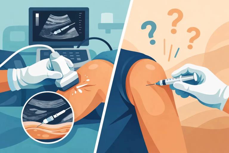
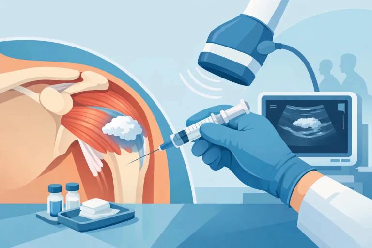
One of the major advantages of ultrasound in a musculoskeletal clinic is its role in guiding certain procedures. If an injection is clinically appropriate, ultrasound can show the needle path and the target structure in real time. This may help the clinician place treatment accurately into or around a joint, bursa, tendon sheath or other indicated area.
Image guidance is not necessary for every injection, and an injection is not automatically the right answer simply because a scan identifies inflammation. The decision should take account of your diagnosis, duration of symptoms, previous treatment, activity goals, medical history and the potential benefits and risks.
For example, rehabilitation may be the main treatment for many tendon problems, while a guided injection may have a role in selected cases such as a painful bursa, inflammatory joint condition or certain nerve entrapments. A careful diagnosis matters because the wrong intervention, or an intervention at the wrong time, may not improve the underlying problem.
What point-of-care ultrasound cannot show
Ultrasound is highly useful, but it has limits. Sound waves do not pass well through bone or air, so ultrasound cannot assess the inside of a joint in the same way as MRI, and it is not the best imaging test for spinal pain. It may also be less informative when a structure lies deep beneath the surface.
An X-ray may be needed to assess fractures, bone alignment or more established arthritic changes. MRI may be more appropriate for suspected internal cartilage injury, deep muscle injuries/pathology, bone marrow concerns, complex ligament injury or when symptoms and ultrasound findings do not match.
Its value lies in using the right test at the right time and avoiding unnecessary delay where ultrasound can answer the clinical question.
Is point-of-care ultrasound safe?
Diagnostic ultrasound uses sound waves rather than ionising radiation. It is generally considered safe when used appropriately by trained clinicians. There is no recovery period after a standard scan, and you can return to work, driving and usual activities straight afterwards.
If you are having an ultrasound-guided injection, your clinician will discuss the specific procedure, expected benefits, alternatives and possible side effects beforehand. Advice after treatment will depend on the area treated and the medication used.
Pain that is persistent, worsening or limiting your work, sleep, mobility or sport deserves a clear assessment. A prompt clinical review with point-of-care ultrasound, where suitable, can turn an uncertain symptom into a focused plan for recovery.